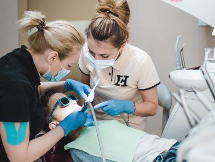
Quote from an American doctor «You cannot treat what you cannot see» especially true for dentists, when dentition visibility is limited by gums, lips, tongue, oral mucosa and soft palate.
The use of optical magnification in dentistry has started in 1981, when an American N. Chivian first applied 8x magnification of tooth picture. In 10 years, Gary B. Carr improved dental microscope. Now the construction could be attached to both the floor and the ceiling. But the main benefit was the possibility to see dentition from different angles under additional backlight and substantial magnification. Since then, the construction of the microscope has been repeatedly improved. Now, dentists can increase the working field by 30 times.
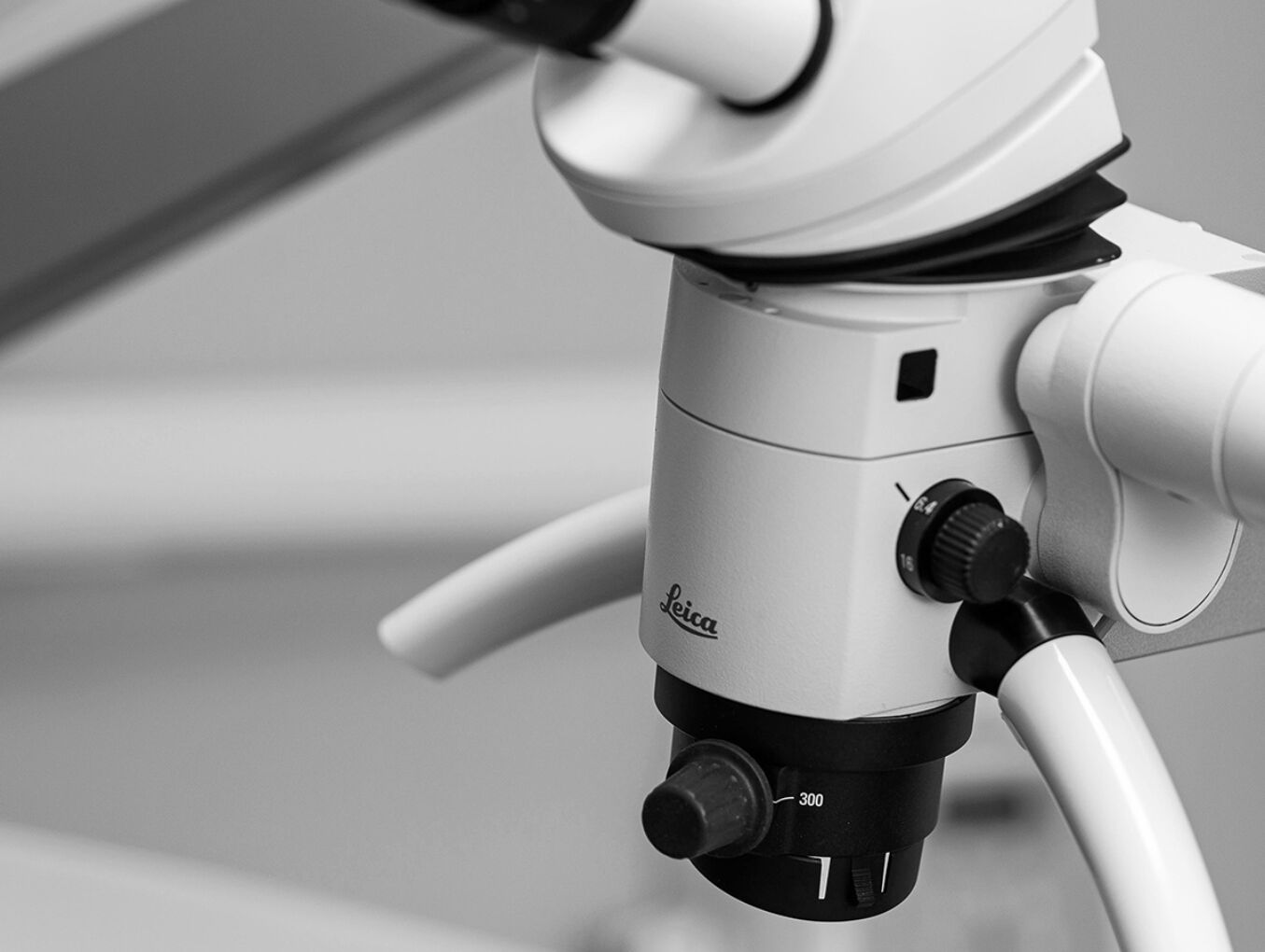
At our clinic we use one of the most modern microscopes Leica M320.
Benefits of microscope usage
- Increased magnification of working area. With the help of a microscope, a doctor has the possibility to evaluate border transition of gums and crowns (or veneers), detect additional canals in a tooth, tooth root cracks, which influences great on tooth prognosis in the future. Insufficiently treated canals lead to repeated inflammation and subsequently to tooth extraction. Furthermore, if you miss and do not see a crack in the root of the tooth, when choosing an orthopedic construction (crown), the patient may have an unpleasant surprise as crown falling out along with a part of tooth.
- Improved work efficiency. With a 30x magnification, the dentist precisely sees the work area. He conducts treatment more thoroughly, which guarantees a high-quality result and reduces the frequency of client visits.
- Identification of the problem of «white» spot in the early stages. «White spot» is a signal of caries (enamel part with demineralization). Through the use of a microscope in a clinic, a doctor with 100% percent probability can diagnose parts of demineralized enamel (flushing out of enamel its components), that further leads to caries. Such early diagnostics is crucially important because it avoids interference with enamel and further filling. At most cases special professional care means can be prescribed, both for home use and treatment course in the clinic. These are special applications, rubbing medicine into tooth enamel, rinse aids.
- Removing the patient’s fear of dental manipulation. If the microscope is connected to the camera and displays an image of the working field on the screen, on patient’s wish, he can see his problem in the oral cavity and all the stages of its solution by the doctor.

What dental work cannot be performed without a microscope?
- Therapy of tooth canals. If no one has previously treated a tooth, then the patient is kind of lucky. The endodontist will immediately determine the exact number of canals, work them through the entire length and thoroughly fill them up.
- Tooth canal filling. When a dentist treats up the canals after previous dentist that worked without a microscope. There is a risk of repeating errors that lead to repeated inflammation: missed canal, choice of wrong movement of the instrument inside a canal, development of a step in a canal, that will further complicates treatment. Work with a microscope allows cleaning the tooth canal from poor-quality fillings, which will further extend the function of the tooth for many years.
- Work with the open root of tooth apex. This is one of the most difficult types of work that is not recommended without a microscope. The removal of the filling mass into the bone tissue (beyond the tip of the tooth) can lead to complications. Constant «leakage» of root canal can also depressurize the filling mass in the canal in the future and cause re-inflammation. Only with a microscope, an endodontist can not only close the tip of the tooth canal, but also fill it with high quality.
- Therapy of cysts and granulomas. Before the use of a microscope teeth with cysts and granulomas were extracted, and the patient subsequently needed prosthetics. Today, such cases are successfully treated with the use of magnifying equipment.
- Teeth restoration. The microscope allows the dentist to examine in detail the enamel cracks, the depth of caries damage, and to clean the area as thoroughly as possible for further application of restoration material.
- Enamel «white spots» diagnostics.
The use of a microscope is a real necessity. Its usage reduces number of visits to a dentist and makes them more effective.
Welcome to our clinic.