
Correct diagnosis makes half the success of treatment.
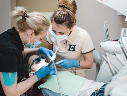
Especially, qualitative diagnostics is important in dentistry, when substantial part of dentist’s working zone is hidden under the gum and visually doctor can evaluate just about 50% of the working area.
X-rays can demonstrate bone and periosteum condition, tumors, evaluate tooth hard tissues. And in this case orthopantomograph became an indispensable assistant of dentists.
Digital image — orthopantomogram simultaneously shows the status of both jaws and adjacent areas (temporomandibular joint, maxillary and other circumnasal sinuses), which allows the use of the diagnostic method not only in dentistry, but also otolaryngology.
Benefits of diagnostics
Among the advantages of modern orthopantomograph Gendex we distinguish:
- image accuracy. The digital image shows the status of two jaws at once. In a classical (targeted) X-ray image, the dentist sees only 2–3 teeth. With orthopantomogram we get much more important information. The doctor can assess the condition of the entire oral cavity (bone thickness, presence of hidden caries, inflammatory processes in the canals)
- image high quality. Digital image — orthopantomogram is more contrast-enhanced. It can be enlarged and viewed at different angles on the computer screen
- safety. With a panoramic image with Gendex the patient receives minimum radiation load (less than with classical X-ray).

When orthopantomogram is needed
We recommend this diagnostic on:
- initial patient examination. To assess dental status and prepare a preliminary treatment plan
- bite adjustments
- planning of wisdom teeth extraction. Often, 8th teeth have anomal (incorrect) location. Lie «on the side», shift toward the oral cavity and closely fit to the neighboring teeth. After taking image, the dentist chooses a tooth removal tactic
- assessment of the periodontal tissues state. In the treatment of periodontitis, it is important to objectively assess the state of bone tissue in which the tooth is located
- treatment of caries. If the patient has many teeth with caries, then the dentist for exact diagnosis and choice of strategy will recommend to make a panoramic image. In this way the doctor will determine the degree and depth of damage to the tooth enamel, and proximity of defect to the pulp, state of teeth canals and quality of previous treatment (if it was held). It is much faster and more safe than taking classical X-ray images for each tooth separately.
- control of teeth growth in children. Orthopantomogram shows the state of the bite, displacement or overlapping of the jaws, abnormal teeth cutting. At early detection of these problems adjustment and correction of bite will last much faster than in adulthood.
Contradictions and safety
Regarding the safety of Gendex X-ray diagnostics use, the procedure is recommended for children from 5 years old. This allows promptly to detect bite formation malfunctions or issues with changing baby teeth to permanent ones.
As for pregnant women, we recommend to refrain from orthopantomography in the first trimester. Pregnancy at the second and third trimester is no longer a contradiction to the examination. For additional safety of the future mother and child special protective aprons and overlays are used.
With the use of modern orthopantomograph with low radiation load, the diagnosis made by a dentist specialist will be accurate, and further treatment — timely and qualitative.
Welcome to our clinic.